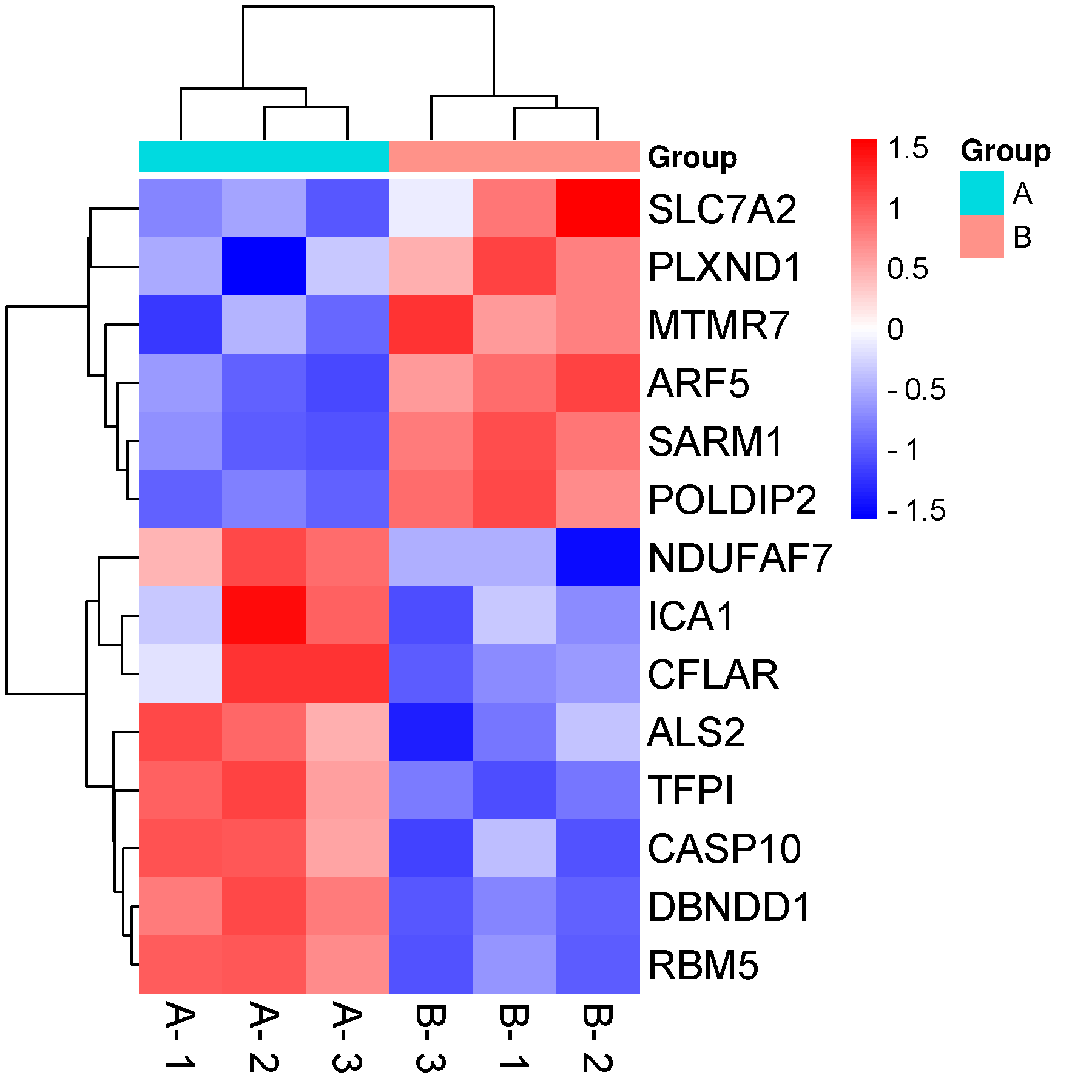
According to cluster analysis in the heat map, STE leads were clustered into two categories, comprising of the right precordial leads (V 1, V 2, V 3) and others (V 4, V 5, V 6, I, II, III, aVF, aVL, aVR). The ST-segment shifts of each lead of each collected ECG could be conveniently visualized in the heat map. STE leads were mainly in the V 1, V 2 and V 3 leads.

In total, 60 cases of electrocardiographic LVH with STE were screened and analyzed. Cluster analysis was carried out based on the heat map and the results were drawn as tree maps (pedigree maps) in the heat map. HemI 1.0 software was used to draw heat maps to display the STE of each lead of each collected ECG. We sequentially collected the electrocardiograms of inpatients in the First Affiliated Hospital of Shantou University Medical College from July 2015 to December 2015 in order to screen cases of LVH with STE.

The aim of this study was to analyze and show data for electrocardiographic left ventricular hypertrophy (LVH) with ST-segment elevation (STE) by a heat map in order to explore the feasibility and clinical value of heat mapping for ECG data visualization. Few papers display ECG data by visual means. Most electrocardiogram (ECG) studies still take advantage of traditional statistical functions, and the results are mostly presented in tables, histograms, and curves.


 0 kommentar(er)
